


Understanding why and how hearing loss happens isn’t easy. After all, the hearing process occurs inside the tiny space of the inner ear. To better study this process, scientists need an improved way to visualize how the components of hearing work. Scientists at Ohio State University used X-ray crystallography, a method that provides images at the molecular level, to look at the makeup of structures called tip links, which are critical to hearing and balance. The scientists built computer simulations based on these high-resolution images to see how tip links respond to sound-generated forces. These models provide important new information about tip link makeup and function to help us better understand a very small, but important, component of our hearing system.
Tip links and the hearing process
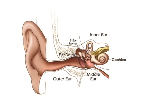
Sound vibrations travel from the middle ear to the inner ear and eventually reach the cochlea, the snail-shaped organ for hearing and balance.
Source: NIH/NIDCD
The hearing process starts when sound vibrations travel from the middle ear to the inner ear and eventually reach the cochlea, the snail-shaped organ for hearing and balance. (Our Journey of Sound to the Brain video explains this process.)
The cochlea is filled with fluid. Sound vibrations make this fluid ripple, creating waves that hair cells inside the cochlea ride as they move. Hair cells are sensory cells inside your ears that are responsible for hearing and balance; they are topped with hair-like structures called stereocilia. Stereocilia are held together by tiny, thread-like strands called tip links, which are made of proteins.
Stereocilia movements stretch the tip links, which in turn open pores (ion channels) to allow positive ions (particles with an electrical charge) to enter the hair cells and convert sound into electrical signals. Our brains then interpret these electrical signals as sound.
What the scientists saw
Scientists knew that tip links were important to hearing and balance, but they did not know how tip links were structured at the atomic level. The high-resolution images in this study showed the scientists how the two proteins that make up tip links connect and work together. The researchers then used supercomputers to build simulations to see the tip links in action.
"If you don’t have the tip link, you can’t hear, and you can’t balance," said Marcos Sotomayor, Ph.D., associate professor of chemistry and biochemistry at Ohio State University and lead author of the study. "[This research] can tell us a great deal more about how these work and what happens when they stop working."
Thanks to high-tech tools such as X-ray crystallography and supercomputers, scientists can now visualize and better understand tip link proteins at the atomic level and further our understanding of how and why some people lose their hearing and balance.
Last Updated Date